From perturbation to evidence
An isolated technique rarely answers a biological question. We design an experimental chain that connects models and perturbations with physiological signals, images, and reproducible analysis.
01 / 04
ManipulateModels and causality
What changes when we alter a physiological condition or a defined cell population?
We generate controlled experimental conditions and modulate specific circuits to test causal relationships among astrocytes, respiratory networks, and disease.
- Experimental context
- Preclinical models of chronic intermittent hypoxia and cortical ischemic stroke.
- Data produced
- Defined perturbations for testing causal relationships in circuits and cell populations.
Capabilities used
- 01Chronic intermittent hypoxiaExperimental model
- 02Cortical ischemic strokeExperimental model
- 03Chemogenetics (DREADDs)Circuit perturbation
02 / 04
MeasurePhysiology in real time
How does an intervention appear in breathing, cardiovascular function, and brain activity?
We record respiratory, cardiovascular, and brain signals in awake animals, aiming to preserve a shared temporal reference across measurements.
- Experimental context
- Awake animals, cardiorespiratory recordings, and 32-channel multichannel LFP.
- Data produced
- Synchronized time series of ventilation, cardiovascular variables, and neuronal activity.
Capabilities used
- 01PlethysmographyRespiratory recording
- 02Cardiorespiratory recordingPhysiological recording
- 03Cardiorespiratory instrumentationExperimental platform
- 04Open EphysOpen platform
- 05Multichannel LFP (32 channels)Electrophysiological recording
03 / 04
VisualizeCells and circuits
Which cells and circuits participate, and what changes in their organization or activity?
We identify and characterize, in vitro, cell populations linked to respiratory circuits while preserving sample, labelling, and acquisition context.
- Experimental context
- Immunofluorescence-labelled tissue and brain slices for in vitro studies.
- Data produced
- Cell distribution, morphology, and calcium signals in defined cells and circuits.
Capabilities used
- 01ImmunofluorescenceTissue labelling
- 02Confocal microscopyStructural imaging
- 03Calcium imaging in brain slicesFunctional imaging
04 / 04
AnalyzeSignals and structure
Which patterns connect an experimental intervention with signals, images, and molecular data?
We transform signals, images, and gene-expression data into reproducible metrics while preserving the relationship between each result and its source record.
- Experimental context
- Electrophysiological signals, images, and gene-expression data.
- Data produced
- Reproducible metrics, temporal relationships, and interpretable multivariate patterns.
Capabilities used
- 01PythonComputational environment
- 02Signal analysisTemporal analysis
- 03Data processingReproducible workflow
- 04Machine learningModelling
- 05Transcriptomic analysisMolecular analysis
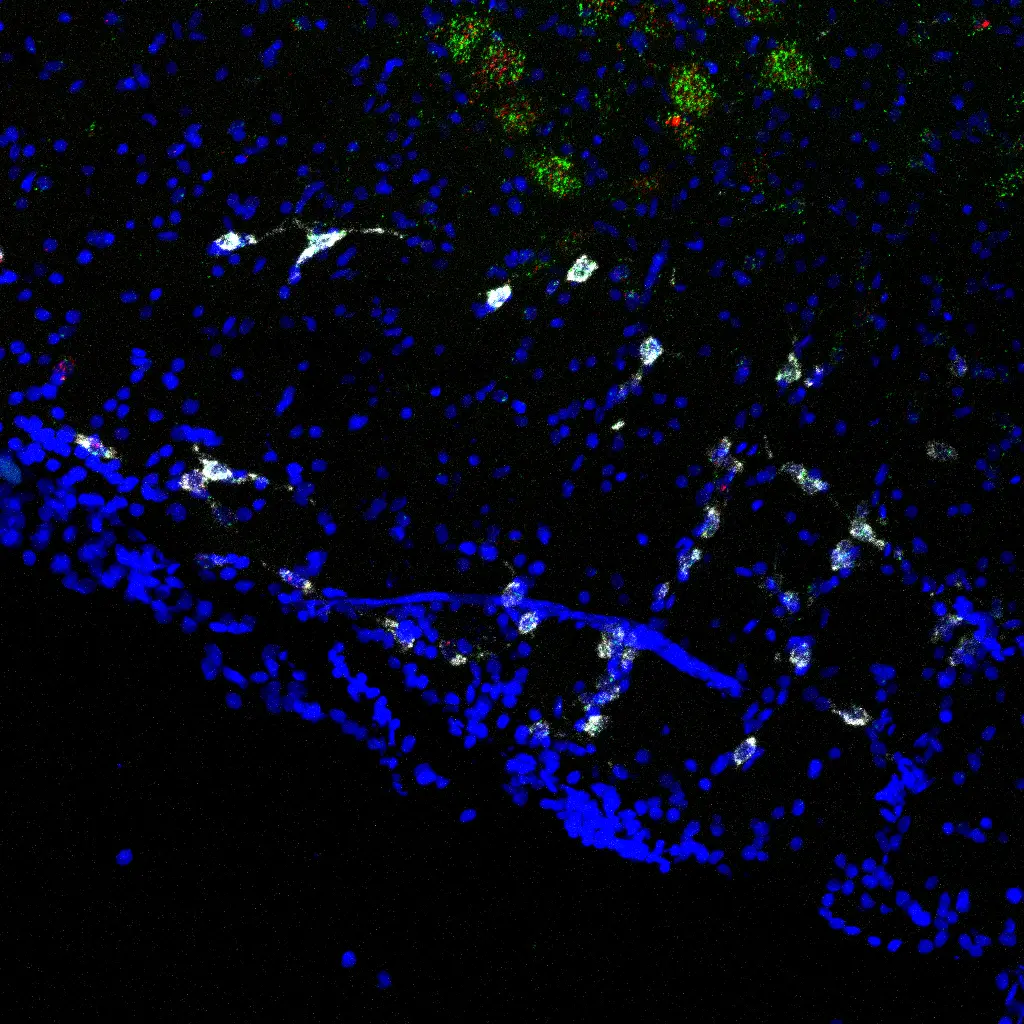
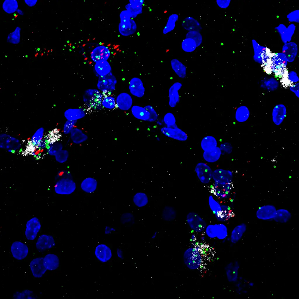
An image is useful only when its context is preserved
Acquisitions are interpreted together with preparation, experimental conditions, channels, and analysis. We therefore distinguish what was observed from what still requires verification.
One chain, not four isolated techniques
Interpretation becomes stronger when a defined perturbation can be followed from physiological recording and imaging to a traceable metric.
Discuss an experimental designEvidence chain
- 01Model or perturbation
- 02Physiological recording
- 03Imaging and tissue
- 04Analysis
- 05Interpretation