Active question · published background
Which neuronal and astrocytic mechanisms sustain breathing rhythm and cardiorespiratory homeostasis?
We study how neurons and astrocytes generate and regulate breathing rhythm and cardiorespiratory homeostasis.
We investigate how brainstem neurons and astrocytes regulate breathing, and how these signals relate to physiology, cognition, and disease.
Institute of Biomedical SciencesHuechuraba · Santiago, Chile
As we sleep, walk, or exercise, the body’s needs change. Brain circuits receive that information and adjust the breathing rhythm without requiring conscious thought.
Part of this work takes place in the brainstem, where neurons and astrocytes participate in networks that generate and modulate breathing. Dr. Esteban Díaz Jara studies how these cells coordinate and how their signals relate to respiratory, cardiovascular, and brain activity.
The laboratory examines what changes during intermittent hypoxia or after ischemic brain injury. To follow the process from the cell to the whole organism, it combines physiology, electrophysiology, microscopy, and data analysis.
How does coordination among cells and respiratory circuits change before disruption becomes visible across the whole organism?
Three concepts for reading this site
From cellular mechanisms to cognition, we study how breathing is generated, how it synchronizes other brain networks, and what changes in disease.
Active question · published background
We study how neurons and astrocytes generate and regulate breathing rhythm and cardiorespiratory homeostasis.
Developing research line
We investigate how respiratory oscillations synchronize cortical and hippocampal circuits.
Previous evidence · new models active
We examine the two-way relationship between brain alterations, respiratory function, physiology, and cognition.
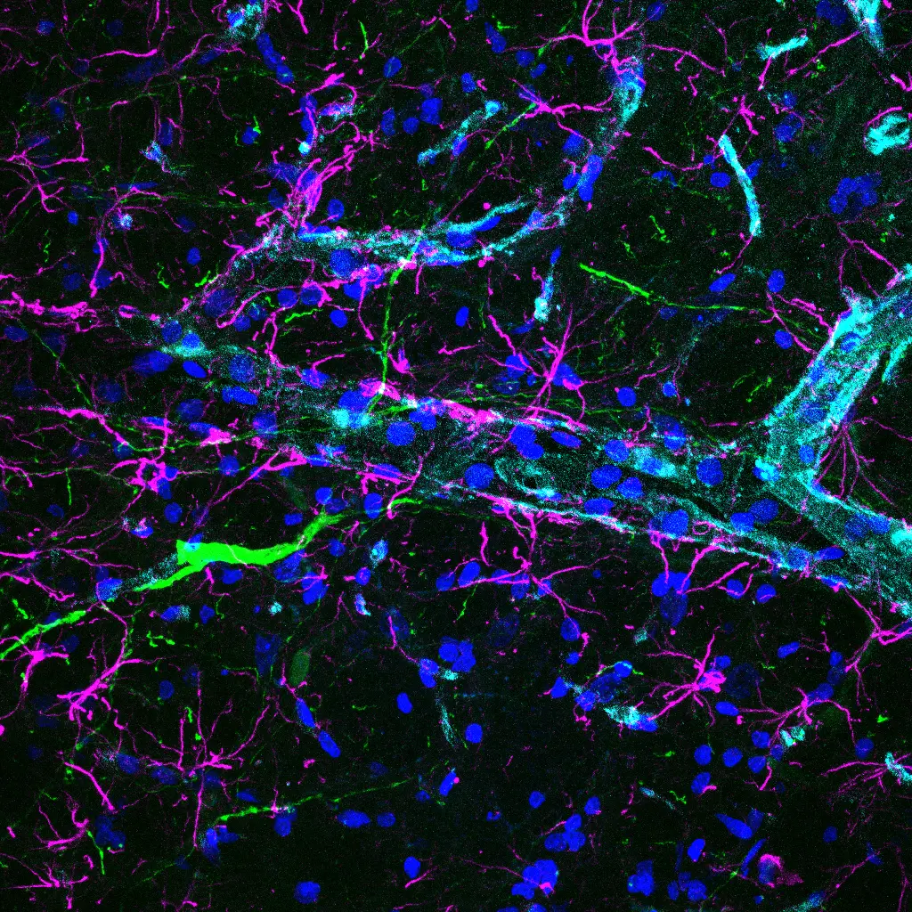
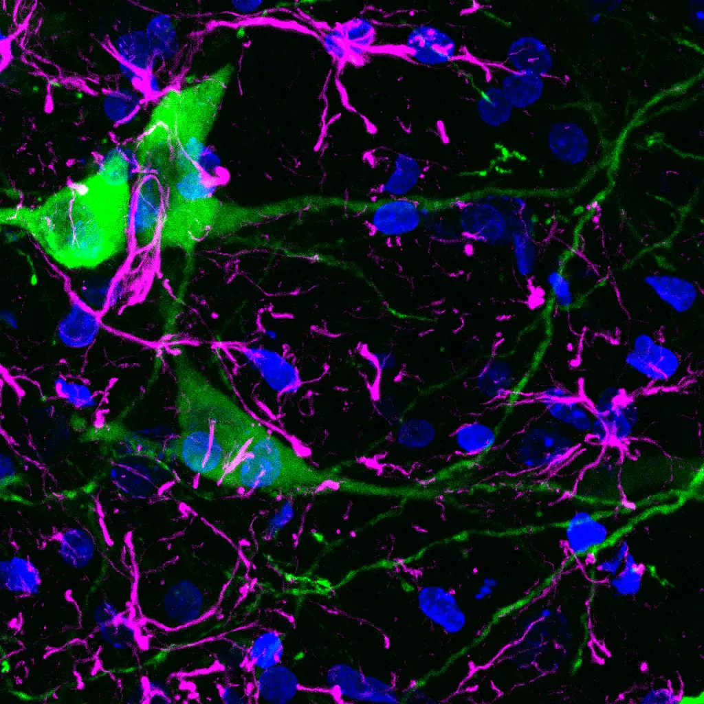
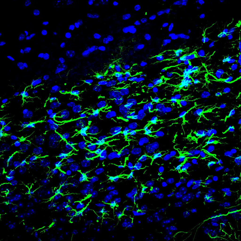
DJL / IMAGE REGISTER / 01–03
These acquisitions are shown in their original channels. Anatomy, markers, and scale are attributed only when the associated experimental record supports them.
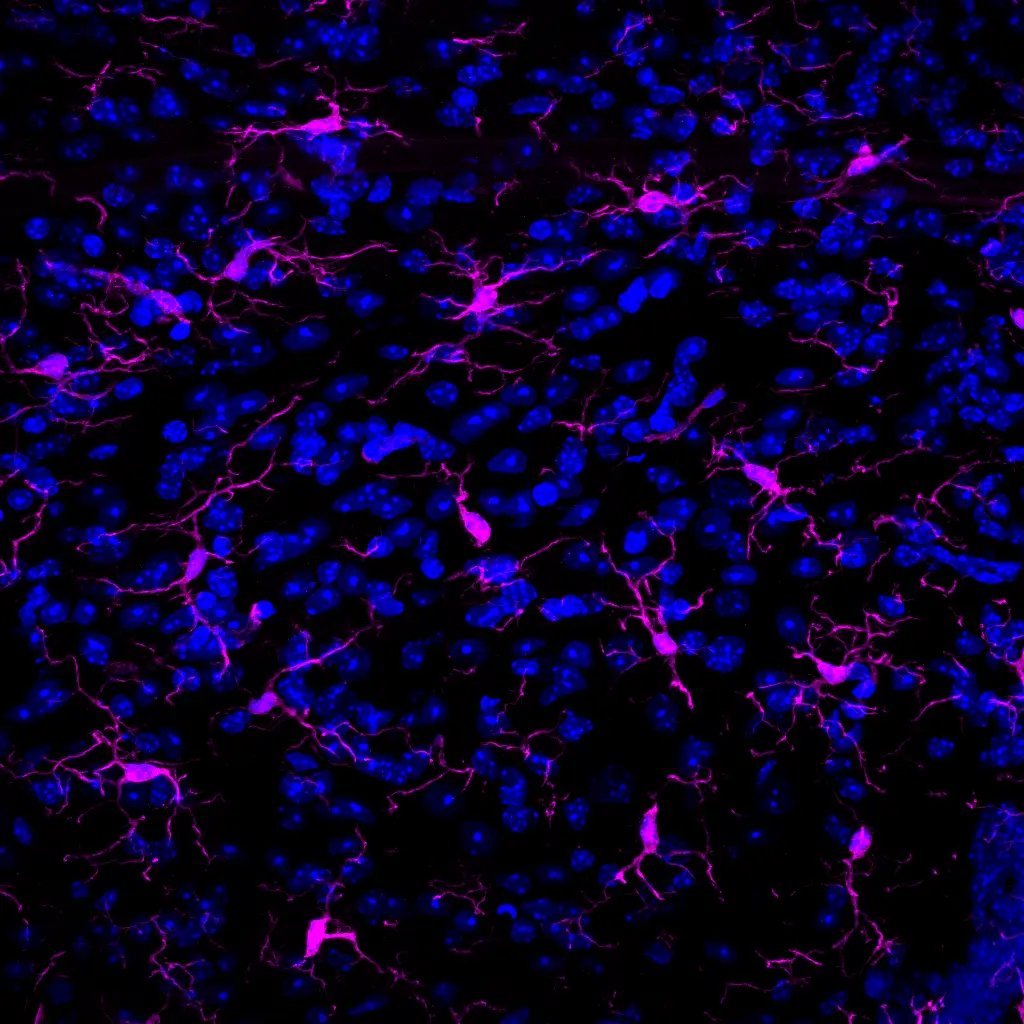
We combine disease models, physiological recordings in awake animals, microscopy, electrophysiology, and computational analysis to connect cellular changes with cardiorespiratory and brain activity.
TechniquesDefined perturbations for testing causal relationships in circuits and cell populations.
Synchronized time series of ventilation, cardiovascular variables, and neuronal activity.
Cell distribution, morphology, and calcium signals in defined cells and circuits.
Reproducible metrics, temporal relationships, and interpretable multivariate patterns.
The program receives funding to investigate neural cardiorespiratory control and astrocyte signaling.
Dr. Esteban Díaz Jara · End date:
Dr. Esteban Díaz Jara · End date:
A selection of recent papers from Esteban Díaz-Jara’s scientific record. Each entry links to its editorial source, while the catalog preserves its bibliographic provenance.
Explore all publicationsKatherin V. Pereyra, Esteban Díaz‐Jara et al.
Katherin V. Pereyra, Alexandra Las Heras et al.
Camilo Toledo, David C. Andrade et al.


Esteban Díaz Jara leads the scientific program; Sinay C. Vicencio Orellana coordinates laboratory operations and contributes to molecular biology, physiology, and microscopy.
View profiles and rolesWe can talk with students and researchers who bring a defined question in respiratory neurophysiology, astrocyte signaling, microscopy, or computational analysis.